


MeCP2 polyclonal antibody
| 货号 | C15310088 | 售价 | 咨询 |
| 规格 | 100 ul | CAS号 |
- 产品简介
- 相关产品
Alternative names: AUTSX3, MRX16, MRX79, MRXS13, MRXSL, PPMX, RTS, RTT
Polyclonal antibody raised in rabbit against human MeCP2 (Methyl-CpG-binding domain protein 2), using 3 different KLH-conjugated synthetic peptides containing an amino acid sequence from the N-terminal, the central and the C-terminal part of the protein, respectively.
| Lot | A131-004 |
|---|---|
| Concentration | not determined |
| Species reactivity | Human |
| Type | Polyclonal |
| Purity | Whole antiserum |
| Host | Rabbit |
| Precautions | This product is for research use only. Not for use in diagnostic or therapeutic procedures. |
| Applications | Suggested dilution | References |
|---|---|---|
| ELISA | 1:100 - 1:500 | Fig 1 |
| Western Blotting | 1:2,000 | Fig 2 |
-
Validation Data
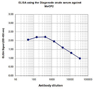
Figure 1. Determination of the titer
To determine the titer, an ELISA was performed using a serial dilution of the Diagenode antibody directed against human MeCP2 (Cat. No. CS-088-100), in antigen coated wells. The wells were coated with the peptide used for immunisation of the rabbit. By plotting the absorbance against the antibody dilution (Figure 1), the titer of the antibody was estimated to be 1:25,000.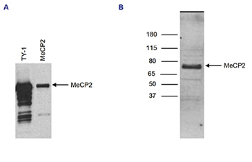
Figure 2. Western blot analysis using the Diagenode antibody directed against MeCP2
Figure 2A: Human osteosarcoma cells (U2OS) were transfected with an expression vector for TY1-tagged MeCP2. The presence of the TY1-MeCP2 in the cell lysates was demonstrated by western blot analysis with the Diagenode antibody against the TY1-tag (Cat. No. MAb-054-050) (lane 1) and with the antibody against MeCP2 (Cat. No. CS-088-100) (lane 2), diluted 1:2,000 in TBST containing 3% milk powder.
Figure 2B: Western blot was performed on nuclear extracts from the human leukemic monocyte lymphoma cell line U937 (60 μg) with the Diagenode antibody against human MeCP2 (Cat. No. CS-088-100), diluted 1:2,000 in TBST containing 3% milk powder. A molecular weight marker (in kDa) is shown on the left. The location of the protein of interest is indicated on the right.